AI-driven medical image and device performance analysis
AI-Supported Medical Image Management and Analysis Software for Clinical Trials
FDA 21 CFR Part 11 compliant imaging platform purpose-built for clinical trials and device evaluation: RAYLYTIC Image Lab is an AI-powered, scalable medical image management and analysis software that harmonizes the capture, storage, exchange, and analysis of medical imaging data.
- Regulatory-ready data
- Designed for sponsors, CROs & clinical research
- Fully compliant
Trusted by top healthcare innovators.


Others sell AI tools.
We use AI-driven image analysis to deliver regulatory-grade evidence at scale.
Unlike single-purpose AI radiology platforms that deliver measurements in isolation, RAYLYTIC Image Lab is a medical image analysis software that delivers reproducible, highly contextual, and fully auditable data.
Every result comes with a transparent, traceable record: who performed the analysis, which tools were used, and how conclusions were reached.
- Regulatory-grade results for submission to FDA and similar international bodies
- Accelerate study timelines through centralized image upload and analysis
- Smart image QC, protocol validation
- Comprehensive audit trails for complete data traceability


Eliminate manual reconciliation across systems.
RAYLYTIC Image Lab brings every medical image, read, and result into a single, unified platform. Sponsors and trial teams no longer juggle multiple systems. Workflows are streamlined, data is instantly traceable, and audit-ready imaging is available whenever it’s needed, reducing risk and accelerating trial timelines.
Drag and drop upload of DICOM files via an integrated DICOM Image Manager
HIPAA and CFR Part 11-compliant browser-based image de-identification
RAYLYTIC Image Lab uses smart image quality control to ensure high image quality, including:
Anatomical coverage
Correct imaging plane (e.g., anteroposterior, lateral)
Compliance of DICOM metadata with trial protocols
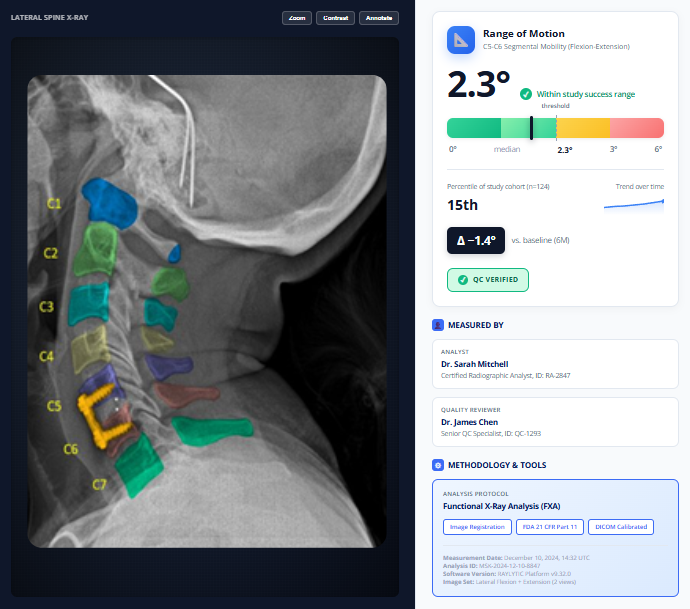
Expert radiologist reviews for qualitative radiographic parameters
Integrated radiological reivew module
Blinded reviews
Adjudication workflows (resolution of discrepancies)
High-precision, AI-based analysis of medical imaging data for regulatory submissions
Automated or semi-automated assessment of over 150 radiographic parameters with reproducible results
- Integrated smart image assessment
- Real-time visibility of data completeness across sites and studies by image type or endpoint
21 CFR Part 11 compliant audit logs
Compliant with GDPR, GCP, and HIPAA
ISO 27001 and ISO 13485 certified
Reliable medical image management and analysis you can trust across your studies.
Reproducible Results Across Studies
Standardized workflows cut variability and ensure imaging measurements stay consistent across sites, subjects, and longitudinal visits.
Built-in Audit Trails
Every measurement and workflow step is tracked automatically, giving you full transparency and confidence for audits and regulatory submissions.
Faster Study Execution
AI-assisted analysis and automated quality control reduce manual review time without sacrificing the rigor your trials require.
Study-Ready Outputs
Results are delivered in standardized, analysis-ready formats that integrate seamlessly into clinical study reporting.
Regulatory-Ready, End-to-End Workflows
Unlike standalone AI tools, RAYLYTIC ensures every step is fully validated, auditable, and compliant, giving you confidence regulators will accept your data.
Trusted by Top Healthcare Innovators
Our platform is designed for sponsors, CROs, and research teams who demand the highest standards for accuracy, traceability, and reproducibility.
AI-driven radiographic measurement & device performance evaluations
AI-driven medical image analysis within RAYLYTIC Image Lab enables consistent, quantitative evaluation of imaging data across studies while meeting the rigor required for clinical research and regulatory use.
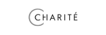
Accurate, Reliable, and Reproducible Spine Imaging Measurements
Conduct rapid and ultra-precise measurements for morphological parameters (Cobb Angle, Lordosis, etc.) and all major types of spinal therapies—including motion preservation, fusion, and deformity correction—and generate reliable, robust, and cost-effective spinal measurements for clinical research.
- High-precision measurements and analysis
- Automated quality control and validation
- Faster research results and reporting
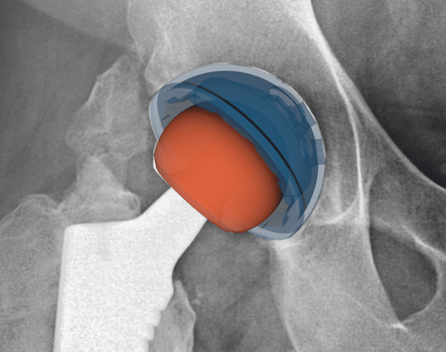
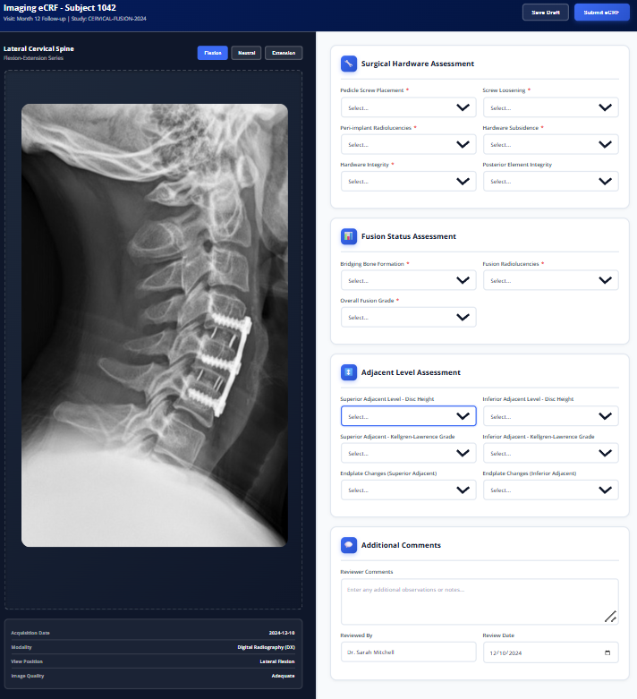
High-Precision Hip Implant Alignment and Performance Analytics
Conduct rapid and ultra-precise measurements for morphological parameters and all major types of hip therapies and generate reliable, robust, and cost-effective measurements for clinical research.
- High-precision measurements and analysis
- Automated quality control and validation
- Faster research results and reporting
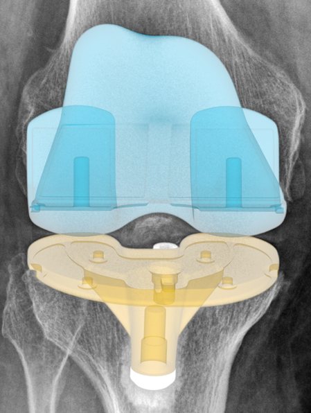
Knee Implant Positioning and Performance Intelligence
RAYLYTIC Image Lab enables high-precision implant positioning and performance such as component alignment, rotation, migration, and wear for faster, more consistent research results.
- High-precision measurements and analysis
- Automated quality control and validation
- Faster research results and reporting
Why RAYLYTIC Image Lab works for every role
RAYLYTIC Image Lab enables sponsors and clinical research organizations to run structured AI image analysis workflows with built-in quality control, traceability, and standardized outputs across the full lifecycle of a clinical study.
For clinical study sponsors
Accelerate your path to approval
- Regulatory-grade data for submission to FDA and international regulatory bodies
- Eliminate inter-observer variability with centralized expert analysis
- Reduce study timelines through parallel processing capabilities
- Comprehensive audit trails for complete data traceability
For Contract research organizations
Scale with confidence
- Seamless integration with existing clinical trial workflows
- Handle multi-site studies with consistent measurement methodology
- Real-time dashboards for study progress monitoring
- Dedicated support from measurement methodology to final report
More than standalone AI models: complete, auditable imaging workflows designed for regulatory submission.
With RAYLYTIC Image Lab, you can ensure every image collected is high-quality, compliant, and ready for analysis across all sites and modalities.
- 21 CFR Part 11 compliant
- Integrated query management
- Real-time access to data, images, and reports
- Imaging eCRFs for independent radiologist review
We use smart image quality control to ensure high image quality, including:
Anatomical coverage
Correct imaging plane (e.g., anteroposterior, lateral)
Compliance of DICOM metadata with trial protocols
Our Proven Process
Five steps to turn imaging data into actionable insights.
Endpoint standardization
Targeted site training
Smart image quality control
AI-driven image analysis
Clear, actionable reports & regulatory-ready datasets
Discover the full list of MSK measurements.
RAYLYTIC Image Lab enables AI-driven image analysis across a wide range of quantitative imaging biomarkers used in clinical trials and medical device studies, supporting consistent, reproducible measurement across modalities, anatomies, and timepoints. Measurements are delivered within regulatory-grade workflows with full traceability and auditability.
Spine
Selected morphoanatomical spine measurements and device performance metrics.
● Spinal Measurements
Angular Range of Motion (RoM, °)
- Schulze M, Trautwein F, Vordemvenne T, Raschke M, Heuer F. A method to perform spinal motion analysis from functional X-ray images. J Biomech. 2011
Most popular
Anterposterior Translational Motion (mm)
- Kelly MJ, Gelfand B, Radcliff K, Mo FF, Felix BA, Babak Kalantar S. Interim 1-Year Radiographic and Clinical Outcomes Following Anterior Cervical Discectomy and Fusion Using Hydroxyapatite-Infused Polyetheretherketone Interbody Cages. Int J Spine Surg. 2024 May 6;18(2):122-129.
Most popular
Cobb Angles
- Berlin C, Adomeit S, Grover P, Dreischarf M, Halm H, Dürr O, Obid P. Novel AI-Based Algorithm for the Automated Computation of Coronal Parameters in Adolescent Idiopathic Scoliosis Patients: A Validation Study on 100 Preoperative Full Spine X-Rays. Global Spine J. 2024 Jul;14(6):1728-1737.
Most popular
Looking for a radiological outcome or MSK measurement not listed here?
We offer over 100 other validated spinal measurements! Contact us to see if we have what you’re looking for or can cooperate on parameter development.
● Device Performance Metrics
Implant Subsidence
Only at RAYLYTIC
Lordotic/Kyphotic Angle Restoration
- Löchel J, Putzier M, Dreischarf M, Grover P, Urinbayev K, Abbas F, Labbus K, Zahn R. Deep learning algorithm for fully automated measurement of sagittal balance in adult spinal deformity. Eur Spine J. 2024
Only at RAYLYTIC
Vertebral Height
Only at RAYLYTIC
Looking for a radiological outcome or MSK measurement not listed here?
We offer over 100 other validated spinal measurements! Contact us to see if we have what you’re looking for or can cooperate on parameter development.
Hip & Pelvis
Quantifies hip joint alignment, implant component positioning, and longitudinal changes with expert-validated, regulatory-grade precision.
● Morphological Measurements
Pelvic Tilt
Only at RAYLYTIC
Acetabular Cup Anteversion & Inclination
Only at RAYLYTIC
Polyethylene (PE) Wear
Only at RAYLYTIC
Looking for a radiological outcome or MSK measurement not listed here?
We offer over 100 other validated spinal measurements! Contact us to see if we have what you’re looking for or can cooperate on parameter development.
● Device Performance & Implant Positioning Metrics
Polyethylene (PE) Wear
- Klebingat S, Bien T, Hürtgen J, Grover P, Dreischarf M, Alkhateeb S, Jäger M, Rose G. Accurate determination of hip implant wear, cup anteversion and inclination through AI automated 2D-3D registration. J Orthop Res. 2023 Sep;41(9):1985-1995.
Only at RAYLYTIC
Hip Stem Migration
- Our migration measurement methodology has undergone rigorous internal validation to ensure clinical-grade accuracy and reproducibility.
Most popular
Radiographic Acetabular Cup Anteversion & Inclination
- Klebingat S, Bien T, Hürtgen J, Grover P, Dreischarf M, Alkhateeb S, Jäger M, Rose G. Accurate determination of hip implant wear, cup anteversion and inclination through AI automated 2D-3D registration. J Orthop Res. 2023 Sep;41(9):1985-1995.
Only at RAYLYTIC
Looking for a radiological outcome or MSK measurement not listed here?
We offer over 100 other validated spinal measurements! Contact us to see if we have what you’re looking for or can cooperate on parameter development.
Knee & Leg
Comprehensive morphological and implant positioning assessments from standard clinical radiographs.
● Morphological Measurements
Femoral tibial angle, medial angle (Hip-Knee-Ankle Angle, mechanical axis °)
Tibial slope angle (°)
Looking for a radiological outcome or MSK measurement not listed here?
We offer over 100 other validated spinal measurements! Contact us to see if we have what you’re looking for or can cooperate on parameter development.
● Device Performance & Implant Positioning Metrics
Implant Component Positioning Assessment from Standard Radiographs
Precise quantification of femoral component rotation, tibial component rotation, and coronal/sagittal alignment: all derived from routine postoperative AP and lateral X-rays.
Published Validation Data
- Coming soon
Only at RAYLYTIC
Polyethylene (PE) Thickness & Wear Behavior
Precise quantification of femoral component rotation, tibial component rotation, and coronal/sagittal alignment: all derived from routine postoperative AP and lateral X-rays.
Published Validation Data
- Coming soon
Only at RAYLYTIC
Tibial component migration
A radiographic measurement that quantifies the displacement of the tibial component relative to the proximal tibia in total knee arthroplasty. Assessed through serial radiographs, migration is measured in both linear (millimeters) and angular (degrees) dimensions across mediolateral (ML) and craniocaudal (CC) axes.
Supporting Literature
- Our migration measurement methodology has undergone rigorous internal validation to ensure clinical-grade accuracy and reproducibility.
Looking for a radiological outcome or MSK measurement not listed here?
We offer over 100 other validated spinal measurements! Contact us to see if we have what you’re looking for or can cooperate on parameter development.
Trusted across the full spectrum of clinical research and innovation.
“With RAYLYTIC we are offering surgeons an intuitive way to generate and access imaging intelligence as part of our clinical registry.”
David Oka
Director of Business Development and Clinical Affairs at Innovasis

2-3x
Reduction in read time compared to manual review methods
<0.2°
Mean error for spine rotational measurements
<1mm
Mean error for spine translational measurements
“The ability to centrally and objectively determine fusion rates in dynamic radiographs was a key factor in the success of our PMCF studies.”
Dr. Stefan Maenz
Head of Clinical Evaluation and Clincial Studies for Aesculap products at B. Braun in Tuttlingen, Germany
Advanced Analytics (UNITY INSIGHTS)
Lorem ipsum dolor sit amet, consectetur adipiscing elit. Ut elit tellus, luctus nec ullamcorper mattis, pulvinar dapibus leo.
Analytics portal
Own your analytics.
Lorem ipsum dolor sit amet, consectetur adipiscing elit. Ut elit tellus, luctus nec ullamcorper mattis, pulvinar dapibus leo.

Ready to elevate your clinical study?
Speak with our team to discuss your specific image review needs and learn how RAYLYTIC can support your research.