Prof. Dr. Jörg Franke // Clinic Magdeburg
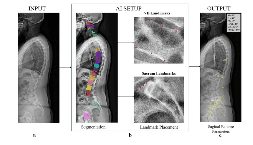
Fully automated method for measuring spinal sagittal balance parameters
Challenge
Measuring parameters of sagittal balance (SB) is essential in the treatment of spinal disorders. However, it is a time-consuming and labor-intensive manual process.
Solution
RAYLYTIC partnered with Prof. Dr. Jörg Franke to develop and systematically evaluate a fully automated algorithm for determining SB parameters.
Outcome
The algorithm demonstrated excellent reliability when compared to manual measurements by experienced physicians. The results of the validation study were published in the European Spine Journal in July 2022.
Excerpt
RAYLYTIC partnered with Prof. Dr. Jörg Franke, chief of orthopedic and emergency surgery at the Clinic Magdeburg in Germany, to develop a fully automated method that would reduce the time and effort needed to compute essential pre- and postoperative SB parameters.
Challenge: Essential, yet time-consuming sagittal balance measurements
Sagittal balance parameters in clinical routines
Reestablishing SB is the goal of many spinal surgeries, as radiographic parameters of SB have been shown to correlate significantly with patient outcomes, like quality of life and condition-specific disability. The accurate pre- and postoperative analysis of SB parameters is therefore essential to the successful treatment of spinal disorders.
For Prof. Franke, global and segmental analysis of these geometric interrelations between spine and pelvis heavily influence his surgical care strategies. Preoperatively, they can be used to guide surgical decision making, while postoperatively, they can serve as a quality metric to determine if surgery achieved and maintained balance in the sagittal plane.
Limitations of current clinical approaches
Current clinical approaches for determining SB parameters are not only time-consuming and labor-intensive, but also rely on the subjective experience of the observer. Although software exists for measuring SB parameters, it still requires manual – and therefore subjective – user input for the placement of anatomical landmarks.
Moreover, higher volumes of imaging data and more complex cases have put additional strain on his staff. According to Prof. Franke, these manual measurements can take up to 15 minutes per patient – a tall order for his clinic’s radiology department that is reckoning with resource shortages.
Taken together, these limitations make the objective and efficient of analysis of SB parameters for different purposes – clinical diagnostics, treatment planning, scientific research, registry and big data analyses – difficult in the face of skyrocketing radiology workloads, which have tripled in the last 15 years.
A fully automated tool for measuring SB parameters could not only alleviate clinics from routine manual tasks, but also elevate patient care by generating insights from massive repositories of data.
Solution: Fully automated algorithm for measuring sagittal balance parameters
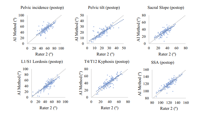
RAYLYTIC partnered with Prof. Franke to develop and systematically evaluate the reliability of an algorithm that could significantly relieve these challenges by automatically computing the following SB parameters:
- pelvic incidence
- sacral slope
- pelvic tilt
- L1/S1 lumbar lordosis
- T4/T12 thoracic kyphosis (TK)
- spino-sacral angle (SSA).
Although recent research has demonstrated the potential of artificial intelligence (AI) to “automatically extract complex radiographic parameters from medical images with high accuracy and precision”1, these studies have conducted validation using small patient cohorts and exclusively preoperative imaging data.
Conversely, the study initiated by RAYLYTIC together with Prof. Franke validated the fully automated algorithm using a larger, 170-patient cohort with as well as without spinal instrumentation. Furthermore, the study differentiated between pre- and post-operative images. Post-operative images are a notorious challenge for AI-based analysis tools due to the difficulty of placing anatomical landmarks in images containing cages, screws, and other instrumentation.
By omitting any specific inclusion criteria and sourcing the images from routine care, it also established a realistic and broad representation of clinical daily routine from the outset.
The algorithm itself is based on two separate convolutional neural networks. The first detects and localizes relevant anatomical structures in the X-ray images, and the second places landmarks on the vertebral bodies, sacrum, and femurs. These can then be used to compute spinopelvic parameters in the sagittal plane.
Outcomes
Excellent reliability
When compared to human-generated manual measurements, the AI-generated measurements demonstrated excellent reliability across the board. The algorithm was able to determine all parameters in 95% of all preoperative images and 91% of all postoperative images with excellent ICC values (PreOp Range: 0.83–0.91, PostOp: 0.72–0.89). Mean errors were smallest for SSA and largest for TK.
Future possibilities
These results are laying the groundwork for the algorithm’s use in clinical routines. As Prof. Franke put it, “Turning deep learning studies such as this one into clinically viable solutions will require validating the algorithm on all the possible parameters of sagittal balance.”
For him, this progress would mean enormous time savings – not only because of the algorithm’s speed, assessing SB parameters in under a minute, but also because AI “is available anytime.” In scientific research, the algorithm shows promise for rapidly analyzing large registry datasets, accelerating the rate at which researchers and clinicians can draw conclusions and correlations.
1 Grover, P., Siebenwirth, J., Caspari, C. et al. Can artificial intelligence support or even replace physicians in measuring sagittal balance? A validation study on preoperative and postoperative full spine images of 170 patients. Eur Spine J 31, 1943–1951 (2022).